Torn Retina Images / Retinal Detachment Nyc Retina Retina Specialists - How is a retinal tear diagnosed?. 13,832 files in 1,208 albums and 49 categories with 72 comments viewed 3,090,757 times A retinal tear can lead to fluid and blood collecting in the eye, which can cause the development of several new floaters and loss of vision if the tear leads to a retinal detachment. Eye flashes can be a symptom of retinal detachment or retinal tears. The diagnosis of a retinal tear requires examination of the retina through a dilated pupil. How does a detached or torn retina affect your vision?
( learn more) a retinal detachment can result in permanent vision loss if it is not treated quickly. The treatment usually takes less than 15 minutes. Warning signs of retinal detachment may include one or all of the following: Eye flashes can be a symptom of retinal detachment or retinal tears. Ophthalmology surgery, pasteur 2 hospital, nice, france, treatment of a retinal detachment through vitrectomy, the doctor is helped by the intern.
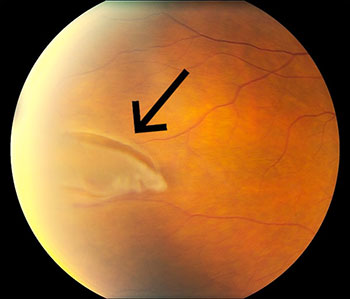
Retinal Tear Causes Symptoms Treatments Assil Eye from assileye.com Symptoms of retinal detachment include an increase in floaters or light flashes in the eye, partial loss of vision, or the visualization of a curtain being pulled over a section of your visual field. The ring of pigmentation (blue arrows) is a reactive repair due to separation of. The retina is the layer of neural tissue that lays across the back of the eye and transforms visible images into signals that are transmitted to the brain for processing. It may be done right in your ophthalmologist's office. ( learn more) a retinal detachment can result in permanent vision loss if it is not treated quickly. Some conditions cause the retina to tear away from the back of the eye. Damage to the retina can cause vision loss and even permanent blindness. Without the delicate retina, there would be no vision.
A detached retina occurs when …
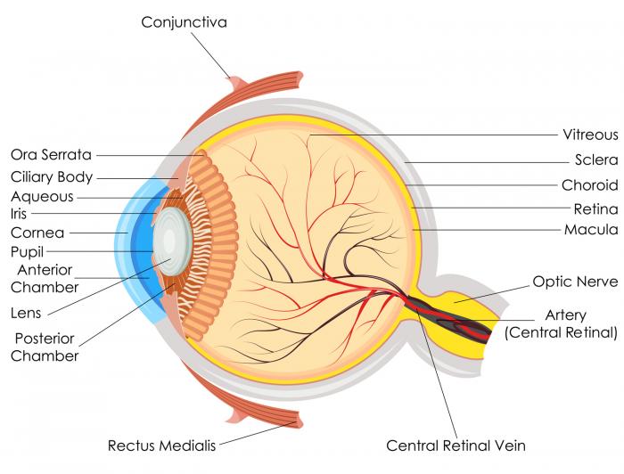
Tears in the retina may impair vision and lead to a detached retina. The vitreous helps to hold the retina in place along the back of the eye. When a retinal tear happens, the vitreous fluid tends to leak through it, causing the retina to be detached from the tissue nourishing it. A related entity are retinal holes. Normally, the retina lies against the back of the eye where blood vessels supply it with oxygen and nutrients. In some cases, a torn retina results in vitreous hemorrhage or bleeding in the transparent cavity of the eye. The term retinal tears and holes may be used interchangeably by some retina specialists. If the tear has led to a retinal detachment, there may be a fixed shadow or dark spot in your visual field. Retinal detachment is a medical emergency. It may be done right in your ophthalmologist's office. Damage to the retina can cause vision loss and even permanent blindness. It occurs when the retina is pushed away by a tumor or due to an accumulation of fluid below the retina due to an inflammatory lesion. Most retinal detachments are a result of a retinal break, hole, or tear.
Retinal holes are typically smaller and have a lower risk for causing a retinal detachment. After images can occur in normal light level settings and resolve in seconds with. A retinal detachment is a separation of the retina from its attachments to its underlying tissue within the eye. There are three types of surgery used to repair a detached retina. Our optic nerves turn these images into electrical impulses, and the brain interprets what we are seeing.
Detached Retina Symptoms Causes Surgery And Treatment from cdn-prod.medicalnewstoday.com How does a detached or torn retina affect your vision? Browse 169 detached retina stock photos and images available, or search for eye exam or glaucoma to find more great stock photos and pictures. Representation of the retinal detatchment, that is detached from the underlying choroid. Contacting an eye specialist (ophthalmologist) right away can help save your vision. The diagnosis of a retinal tear requires examination of the retina through a dilated pupil. Retina's outlying parts help in with the peripheral vision. This is a series of images taken on a patient with a giant retinal tear. The vitreous helps to hold the retina in place along the back of the eye.
Retinal detachment can occur at any age, but it most commonly occurs in caucasian males over the age of 40.
Learn more about the types, causes, risk factors, symptoms, diagnosis, treatment. The term retinal tears and holes may be used interchangeably by some retina specialists. If you have symptoms of a detached retina, it's important to go to your eye doctor or the emergency room right away. Damage to the retina can cause vision loss and even permanent blindness. In front of the retina is the vitreous, a gelatinous type of substance that is made mostly of collagen. Without the delicate retina, there would be no vision. Exudative or solid retinal tear; It may be done right in your ophthalmologist's office. Exudative or solid retinal detachment: Eye flashes can be a symptom of retinal detachment or retinal tears. Browse 169 detached retina stock photos and images available, or search for eye exam or glaucoma to find more great stock photos and pictures. Ophthalmology surgery, pasteur 2 hospital, nice, france, treatment of a retinal detachment through vitrectomy, the doctor is helped by the intern. A detached retina occurs when …
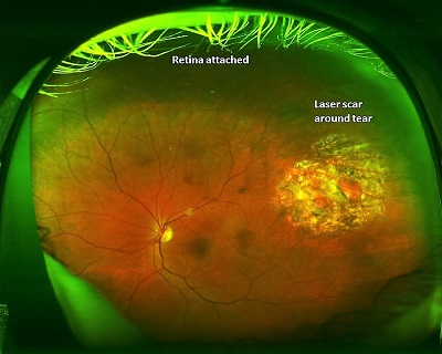
The goal is to keep fluid from going through the tear and detaching the retina. Ophthalmology surgery, pasteur 2 hospital, nice, france. These are serious conditions that can damage your sight. Retinal detachment can occur at any age, but it most commonly occurs in caucasian males over the age of 40. Once the retina has torn, liquid from the vitreous gel (clear gel that fills most of the inside of the eye) passes through the.
Floaters Retinal Tears And Retinal Detachments Visionaware from network.aphconnectcenter.org It may be done right in your ophthalmologist's office. Giant retinal tear, retinal detachment The retina plays a vital role in vision. The diagnosis of a retinal tear requires examination of the retina through a dilated pupil. 13,832 files in 1,208 albums and 49 categories with 72 comments viewed 3,090,757 times When a retinal tear happens, the vitreous fluid tends to leak through it, causing the retina to be detached from the tissue nourishing it. The goal is to keep fluid from going through the tear and detaching the retina. Your ophthalmologist puts a lens on the front of your eye to focus the laser.
There are three types of surgery used to repair a detached retina.
Ophthalmology surgery, pasteur 2 hospital, nice, france, treatment of a retinal detachment through vitrectomy, the doctor is helped by the intern. It's a big day for today's savannah guthrie! A retinal detachment is a separation of the retina from its attachments to its underlying tissue within the eye. A related entity are retinal holes. The recovery timeline is different for each, but the overall range is two to six weeks. The detachment happens when the retina pulls away from its normal position. Retinal tears develop when the vitreous pulls on the retina while retinal holes develop due to progressive thinning of the retina. If the torn retina is associated with some bleeding, your vision may become hazy; What follows is a pictorial, instructive guide depicting and describing various types of retinal holes and tears, their possible etiologies and management strategies. In front of the retina is the vitreous, a gelatinous type of substance that is made mostly of collagen. Symptoms of retinal detachment include an increase in floaters or light flashes in the eye, partial loss of vision, or the visualization of a curtain being pulled over a section of your visual field. A retinal tear is a break in the retina. Our optic nerves turn these images into electrical impulses, and the brain interprets what we are seeing.


0 Komentar